Behavioral Neuroscience
Summers
Crayfish Neuroanatomy
Parallel Gating of Crayfish Escape
Sensory Stimulation of Tail Flip
Parallel Gating of Crayfish Escape
5-HT
Crayfish figures
Escape Neurocircuitry
end Acronyms/Abbreviations
 Neuron types. (a) Multipolar, isopolar neurons (primitive invertebrate). (b) Unipolar, heteropolar neurons (common invertebrate); arrow indicates direction of information flow. (c) Bipolar neuron (common invertebrate). (d) Multipolar, heteropolar neuron (common vertebrate, advanced invertebrate); arrow indicates direction of information flow. (e) Ganglion of unipolar neurons in crayfish. The unipolar cell bodies, surrounded by connective tissue and glia, form a rind around a central neuropile composed of the cell processes (axons and dendrites). Nerve tracts enter or leave the neuropile. Synaptic interactions and integration are carried out in the neuropile. (After G. Retzius, Zur Kenntniss des Nervensystems der Crustacean, Biol. Untersuch., NF 1:–50, 1980)
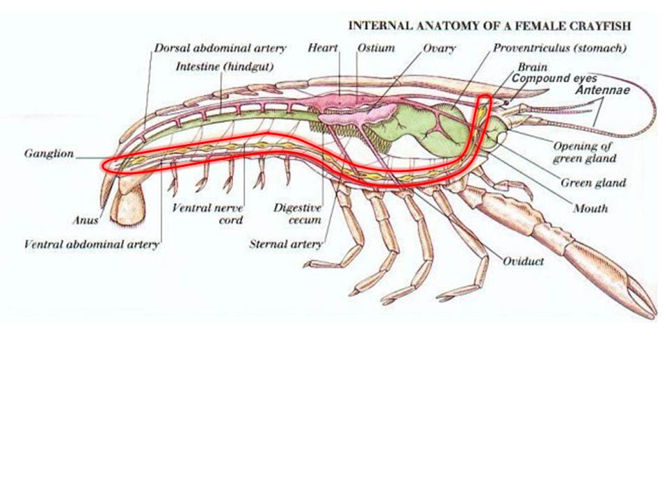
Advanced invertebrates as represented by the crustacean nervous system. (a) Crayfish central nervous system (after W. C. Curtis and J. and J. M. Guthrie, Textbook of General Zoology, 3d ed., John Wiley and Sons, 1938). (b) Photomicrograph of cross section of ventral nerve cord in abdomen of crayfish made near the level of the third root (arrow in a); the largest axons near the top are giant fibers used in escape responses (after J. D. Robertson, Ultrastructure of excitable membranes of the crayfish median-giant synapse, Ann. N.Y. Acad. Sci., 94:339–389, 1961). (c) Relationship between lateral giant and motor giant axons as they make electrical synaptic contact near the third root of an abdominal ganglion in the crayfish (after E. J. Furshpan and D. D. Potter, Transmission at the giant motor synapses of the crayfish, J. Physiol., 145:289–325, 1959).
Neuron types. (a) Multipolar, isopolar neurons (primitive invertebrate). (b) Unipolar, heteropolar neurons (common invertebrate); arrow indicates direction of information flow. (c) Bipolar neuron (common invertebrate). (d) Multipolar, heteropolar neuron (common vertebrate, advanced invertebrate); arrow indicates direction of information flow. (e) Ganglion of unipolar neurons in crayfish. The unipolar cell bodies, surrounded by connective tissue and glia, form a rind around a central neuropile composed of the cell processes (axons and dendrites). Nerve tracts enter or leave the neuropile. Synaptic interactions and integration are carried out in the neuropile. (After G. Retzius, Zur Kenntniss des Nervensystems der Crustacean, Biol. Untersuch., NF 1:–50, 1980)
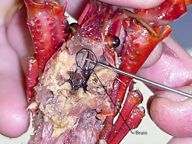
Advanced invertebrates as represented by the crustacean nervous system. (a) Crayfish central nervous system (after W. C. Curtis and J. and J. M. Guthrie, Textbook of General Zoology, 3d ed., John Wiley and Sons, 1938). (b) Photomicrograph of cross section of ventral nerve cord in abdomen of crayfish made near the level of the third root (arrow in a); the largest axons near the top are giant fibers used in escape responses (after J. D. Robertson, Ultrastructure of excitable membranes of the crayfish median-giant synapse, Ann. N.Y. Acad. Sci., 94:339–389, 1961). (c) Relationship between lateral giant and motor giant axons as they make electrical synaptic contact near the third root of an abdominal ganglion in the crayfish (after E. J. Furshpan and D. D. Potter, Transmission at the giant motor synapses of the crayfish, J. Physiol., 145:289–325, 1959).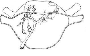 lateral giant interneuron of crayfish anterior view.

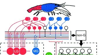
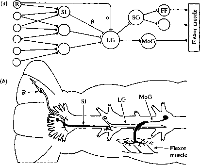 Neuronal circuit for startle behaviour mediated by the lateral giant interneuron. (a) Schematic representation of the excitatory pathway from the mechanoreceptors to the flexor muscles, showing chemical (— and electrical (—|) synapses. Labelled circles represent: the receptors (R); sensory interneurons (SI); lateral giant (LG); segmental giant (SG); motor giant (MoG) and fast flexor motor neurons (FF). The sensory pathways that generate the two components of the compound EPSP (Fig. 3.3c) are labelled a and p. (b) Diagrammatic representation of the arrangement of the above components within the abdominal nervous system. The various components are not drawn to scale and only one segment of the lateral giant is shown; the medial giant and extensor motor neurons are not included. (Modified after Wine & Krasne, 1982).