Yawning
Yawning Animals
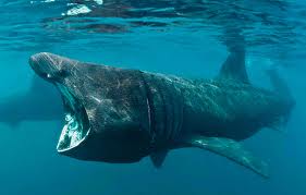
Fish
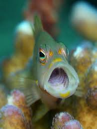
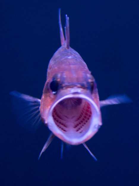
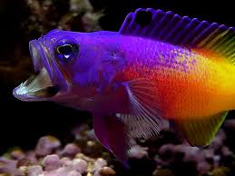 probably not
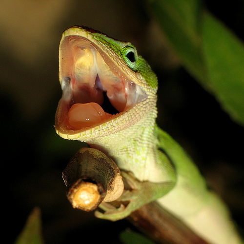
 Reptiles
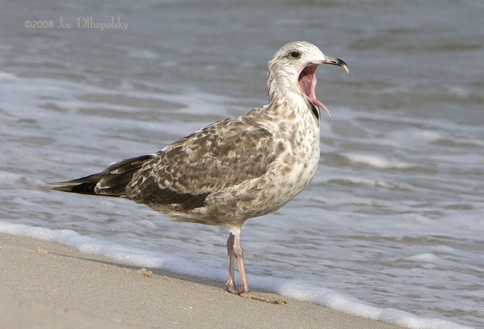
 Birds
Birds
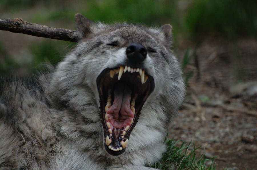
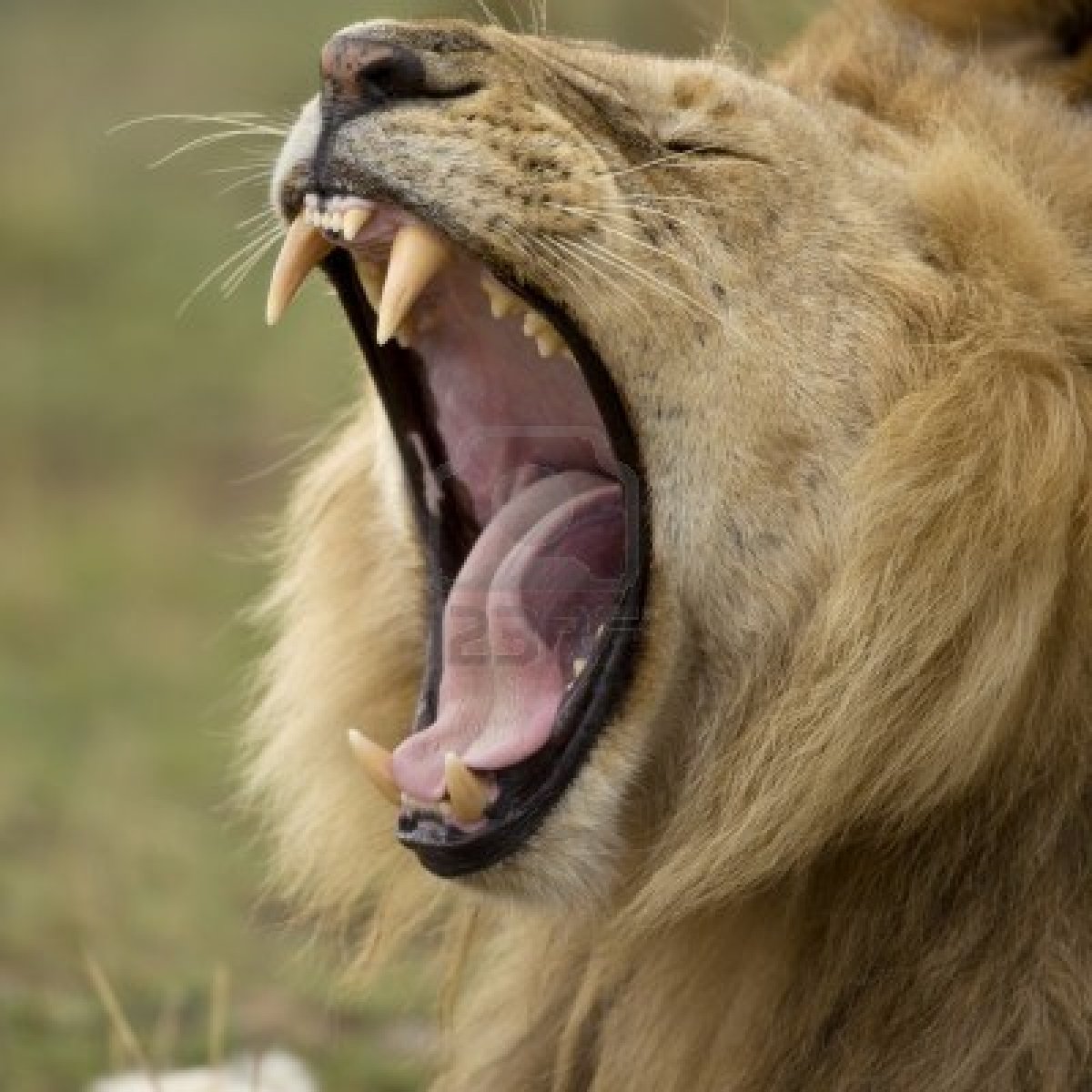
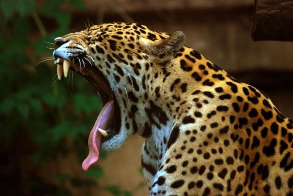
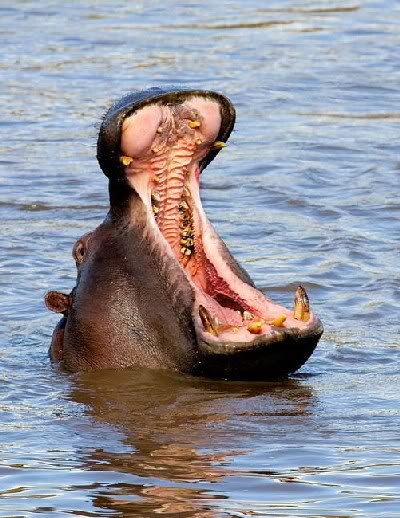
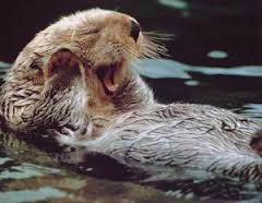
 Mammals


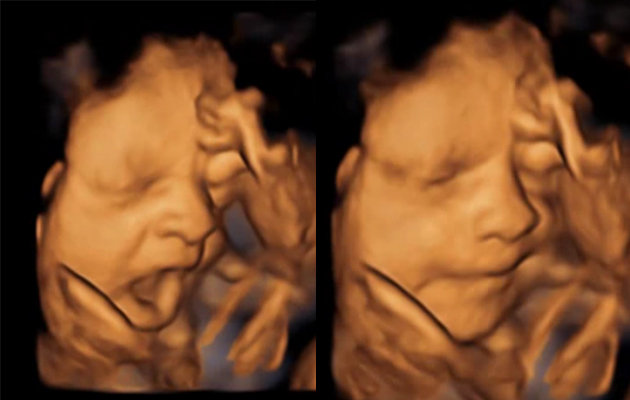
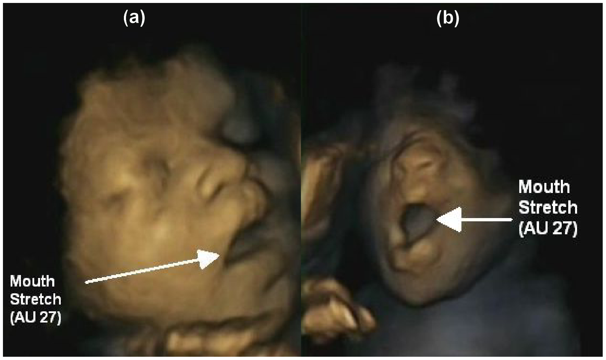
Before birth
Stages of Yawning
Can you tell: (1) Initiation, (2) Acme, (3) Conclusion
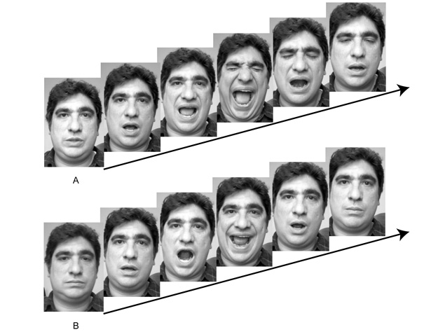

 Look for (1) Initiation: opening the mouth widely
(2) Acme: thorax reaches maximum capacity,
Look for (1) Initiation: opening the mouth widely
(2) Acme: thorax reaches maximum capacity, 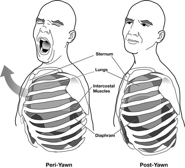 breathing stops, eyes close, Pandiculation
(3) Conclusion: short expiration
breathing stops, eyes close, Pandiculation
(3) Conclusion: short expiration
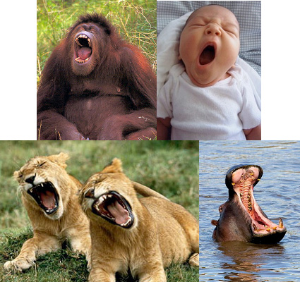
Yawn Contagion
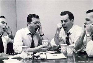
 CONTAGION-YAWN-O-METER
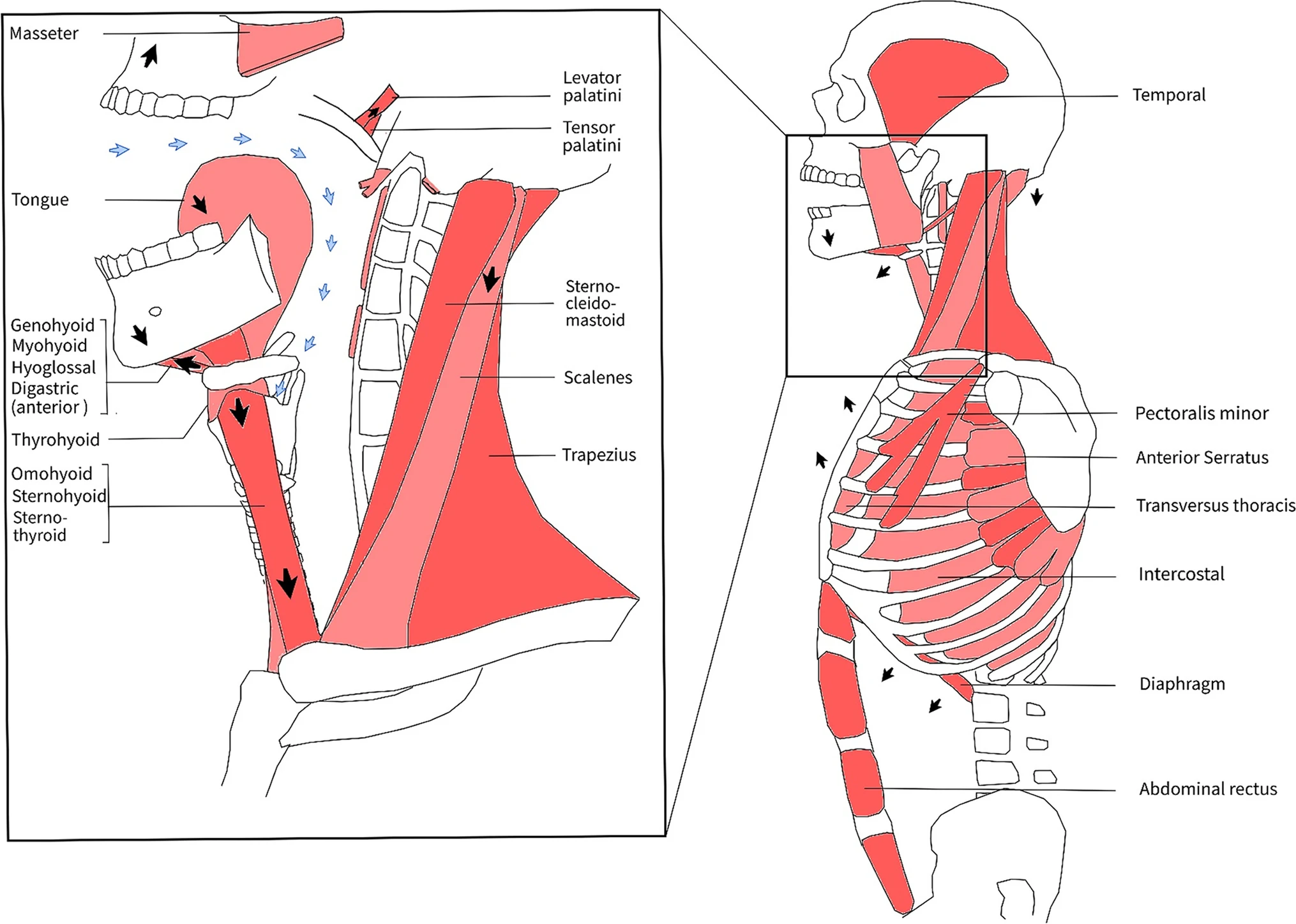 Muscle movements during a yawn: anatomical overview. A yawn can be divided in three phases: (1) the inspiratory phase in which the respiratory muscles gradually contract, (2) the climax (or acme) phase with maximal muscle stretching, and (3) the expiratory phase with muscle relaxation and a satisfied sensation. During the first phase, the subhyoidal muscles (thyrohyoid, sternohyoid, sternothyroid, omohyoid) and floor of mouth muscles (geniohyoid, mylohyoid, hyoglossus, and anterior bellies of the digastric) gradually contract, resulting in jaw opening and pharyngeal dilation (3–4 × diameter). Thereby, the diaphragm, intercostals, and additional respiratory muscles (e.g., scalene muscles, minor pectoral) contract causing deep inspiration (blue arrows). Peak forces are achieved during the second phase, where jaw (e.g., masseter and temporal), pharyngeal (Fig. 3), and other body muscles (e.g., arms, trunk) are powerfully stretched (Fig. 3). Afterwards, prolonged maximal dilation and inspiration muscle tension are released and expiration follows (third phase)
 2D model of muscle movements during a yawn: contracting muscles (red) and stretching muscles (blue). During a yawn, the airway is fully dilated, most prominently in the pharynx. Maximal mouth gaping and pharyngeal widening are achieved by movement of the mandible (A), hyoid (B), larynx (C), and soft palate (D). This results in powerful stretching of jaw and pharyngeal muscles, which are essential for chewing and swallowing. The styloid process (E) and mastoid process (F) are anchor points for swallowing muscles. The stretched muscles include the temporal (1), masseter (2), palatoglossal (3), styloglossus (4), superior pharyngeal constrictor (5), stylohyoid (6), posterior belly of digastric (7), and middle pharyngeal constrictor muscle (8). The palatopharyngeal, stylopharyngeus, and inferior pharyngeal constrictor muscles are stretched as well, however not illustrated here. Stretching is achieved by contraction of antagonist muscles in three groups (Fig. 1): the floor of the mouth muscles (geniohyoid, mylohyoid, hyoglossus, and anterior bellies of the digastric), the infrahyoidal muscles (omohyoid, thyrohyoid, sternohyoid, and sternothyroid), and the pharyngeal and palatal elevators (tensor and levator veli palatine). Stretching muscles is essential to prevent tightening and loss of range of motion. According to our hypothesis, the airway is widened by a yawn and long-term oxygenation is safeguarded