Behavioral Neuroscience
Summers
Sensory input for Rhythmicity
Afferent path to the SCN
Figures of Rhythmicity
Retina-RGC-SCN
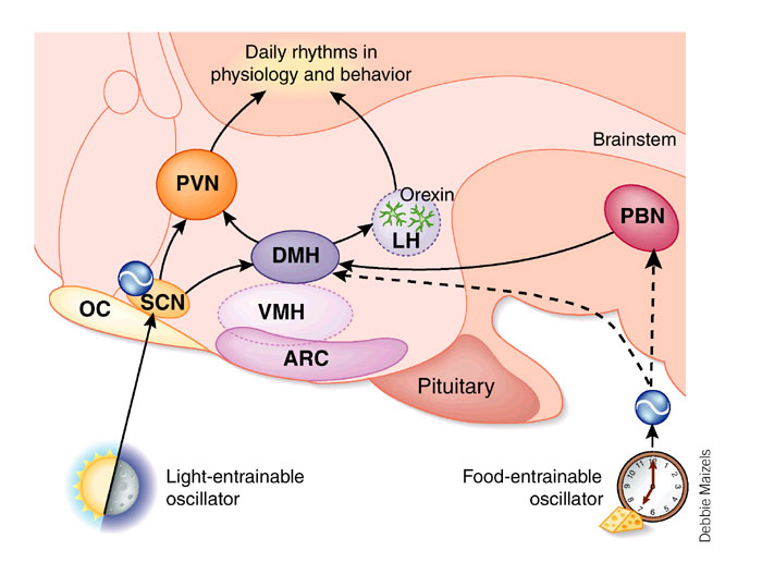
Efferent SCN output
Integration of Rhythms into Behavior
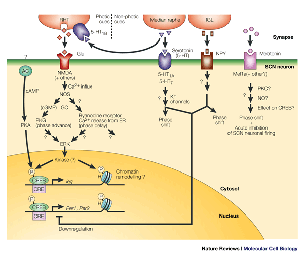
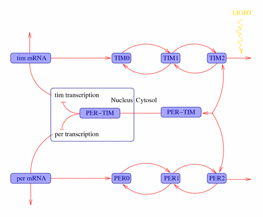
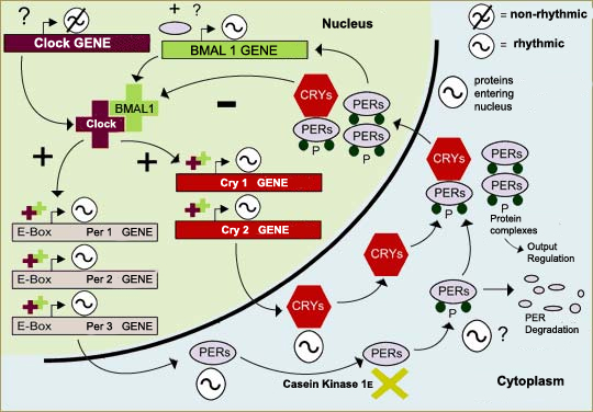
Molecular SCN
end Acronyms/Abbreviations Syllabus
VIP
AVP
GABA
5-HT
 the Brain from Top to Bottom